Imagine you’re trying to find your way through a sprawling, densely populated metropolis and the map you’re using shows only major landmarks and thoroughfares. Back alleys, footpaths, even some of the smallest streets don’t appear. In some cases, entire neighbourhoods are missing.
That’s what it’s like for scientists and medical practitioners trying to navigate the cellular landscape of the human body. There are a staggering 37 trillion cells in the average human. And there’s a lot we still don’t know about them. Many of our cells have never been identified, mapped or even fully understood in terms of the role they play in a healthy body.
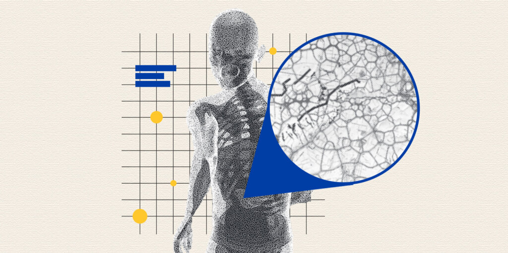
But what if researchers could zoom closer and peer into the human body at a scale that reveals every one of those trillions of cells in fine-grained, vivid detail? An ambitious international effort called the Human Cell Atlas aims to make that possible.
Thousands of researchers around the world are contributing cell samples and analytical data in an attempt to collectively map the location and functions of each cell in the body. So far, about 3,000 participants from 94 countries have signed on to the project, which uses new technology known as single-cell RNA sequencing to tease apart individual cells and study their genetic material.
Traditional research has focused on identifying the genes responsible for a disease, but it hasn’t been able to determine where in the body the gene acts, says Gary Bader, a molecular geneticist at the University of Toronto and one of the coordinators of the atlas’s liver map. Single-cell RNA sequencing enables scientists to pinpoint the type and location of the implicated cells and map them, bridging a critical knowledge gap that will be transformative, he says.
The atlas will serve as a reference tool that will both enhance the success of existing treatments and pave the way for biomedical advances, says Bader. “We will be able to study diseases precisely where they act in the body and develop new treatments for many of them.”
In terms of scope, the cell initiative is bigger than the Human Genome Project, which in the early 2000s undertook the significant challenge of mapping all the genes in a fully sequenced genome. The genome has relatively fewer component parts — only about 20,000 genes. By comparison, since the start of the Human Cell Atlas project in 2016, participants have analyzed more than 121 million cells — and are aiming for billions more. While that number may sound formidable, the pace of the research is accelerating as more scientists join the consortium. (Five years ago, only about 500 were participating.) They’re also aiming for more diversity in terms of the researchers involved and the samples collected to ensure the atlas reflects a broad range of ethnic and geographic groups.
“We want the map to represent humanity as a whole,” says Bader. “It’s important that scientists from all over the world — Latin America and Asia and Africa — are contributing.”
Participants work in networks, each of which focuses on a specific organ or system within the body. Last month, representatives from each of the networks met to discuss their progress in Toronto, where scientists are mapping out the liver.
Bader and two University of Toronto colleagues, immunologist Sonya McParland and transplant surgeon Ian McGilvray, produced a preliminary map of the organ in 2018. The collaborative effort drew on the work of a cross-disciplinary team of doctors and scientists connected through the university’s Medicine by Design initiative, which receives funding from the Canada First Research Excellence Fund. Bader describes the map as a “first draft,” based on an analysis of about 10,000 cells collected from five healthy deceased donor livers. Since then, the network has added considerable data. It has now mapped more than a million cells from a broader cross-section of donors — about 130 samples contributed by labs around the world.
“We still don’t know what many of the cells in the body do,” says Bader. “Learning more about them is what makes this project so exciting.”
Before single-cell RNA-sequencing was developed, researchers applied a “fruit salad” approach to cell analysis, says MacParland, a senior scientist in the Ajmera Transplant Centre at UHN and an associate professor in the University of Toronto’s department of Laboratory Medicine and Pathobiology and the department of Immunology. They knew the liver contained different cell types, she says, but they couldn’t investigate what individual cells did or how they interacted with each other.
The new sequencing technique was developed in 2009 and has been refined over the past decade. It enables researchers to identify the properties of individual cells — break down the fruit salad’s ingredients, in effect, and consider what each item contributes. Just as even a small handful of blueberries can alter the overall taste, small populations of cells can play key roles in liver function, by “driving disease or treating it,” MacParland says. “So rather than looking at averages of how all the cells behave together, it’s important to understand each small population.”
A new cell type discovered by the liver network offers a case in point. The researchers knew the liver contained a population of cells known as macrophages, which perform a debris-clearing function in the organ. But single-cell RNA sequencing revealed that the macrophage population actually consists of two subtypes, one of which promotes inflammation, which can cause complications for patients with liver disease — and in the case of transplants, lead to organ rejection. Armed with this knowledge, medical scientists can develop more effective treatments. Transplant programs can explore ways to suppress pro-inflammatory macrophages, for instance, and “dial up” the other subpopulation, which performs an anti-inflammatory role. “That could make a real difference to the success of a liver transplant.”
Other networks within the consortium are making similarly exciting discoveries. The lung and airways group publicly released its first map in June , based on more than 2.4 million cells from 486 individuals. Its work has also identified some rare and previously unknown cell types, including one that has answered a critical — and longstanding — question about cystic fibrosis. Medical scientists at Toronto’s Hospital for Sick Children discovered the gene that causes the fatal genetic disease in 1989 , but decades of study failed to identify where in the lungs the gene was located. In 2018, cell atlas researchers discovered the main cell type that harbours the cystic fibrosis gene. Further study could lead to new treatments for the disease.
Aviv Regev, then at the Broad Institute of MIT and Harvard, and Sarah Teichmann of the Wellcome Sanger Institute founded the atlas in 2016 as an open-science initiative. And for many scientists, that is one of the most exciting aspects of the cell atlas. Data is regularly uploaded and made available to all researchers, even though the maps are still works in progress.
Open-science proponent Aled Edwards, who established the Structural Genomics Consortium based on similar principles, says that’s a critical part of its success, since it allows a broader base of scientists to consider the findings and accelerate new discoveries. “This is a foundational catalogue — a book that everyone will read, so the last thing you want to do is hide pages,” Edwards says. “You get far more amplification of knowledge if it’s shared by everybody. You can never imagine what ideas some young scientist in another part of the world will have when they see the data.”
There’s no hard deadline for completion of all the maps. And at least in part, that’s because there’s no clear definition of what completion even means. “Science doesn’t have an endpoint,” says Bader. “Once we discover something we didn’t know existed, we say, ‘okay what does this do?’ And then we discover more.”
New technology is always facilitating discoveries, Bader says, and the collaborative, global nature of the cell atlas likely will, too. “As we learn more, we may decide that we need a separate map for Sub-Saharan Africa or for Europe. We’ll likely want maps of diseased livers. And pediatric livers. A liver map for men and a liver map for women. It’s going to be hard to know where exactly to stop.”
MaRS believes “innovation” means advancing Canadian technology for the benefit of all people. Join our mission.
Photo illustration by Monica Guan, Image source: unsplash
This website uses cookies to save your preferences, and track popular pages. Cookies ensure we do not require visitors to register, login, or share any identity information.